
Gum Grafting: Types, Recovery, and What to Expect

If a periodontist has recommended a gum graft, you probably want two things: a clear picture of which graft technique is being proposed for your situation and a realistic week-by-week sense of what recovery actually looks like. The internet tends to deliver one of two extremes. Some sources make grafting sound trivial, and others make it sound far worse than the procedure typically is for most patients. This guide walks through the four main graft types used today, what the donor and recipient sites look like during healing, what to expect for pain and diet across the first month, and what the published evidence indicates about long-term success rates. Dr. Praveen Parachuru performs gum grafting at Prosper Periodontics for patients from across the North Dallas area.
What Are the Four Main Types of Gum Grafts Used Today?
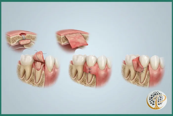
The four primary techniques are the free gingival graft, the connective tissue graft (the most commonly used and considered the gold standard), the pedicle graft, and the acellular dermal matrix graft using donor tissue, and each is selected based on the recession pattern, the amount of attached gum tissue present, and the cosmetic demands of the area.
The connective tissue graft (CTG) is the workhorse procedure for treating recession on visible teeth. A small flap is elevated on the palate and a section of subepithelial connective tissue is harvested from beneath that flap. The harvested tissue is placed under a partial-thickness flap created at the recipient site over the receded root, and both sites are sutured. Because the tissue at the recipient site is covered by the patient’s own gum tissue, the cosmetic blend is typically excellent. The CTG carries the strongest published track record for root coverage in single and multiple recession defects.
The free gingival graft (FGG) takes a full-thickness piece of palatal tissue (epithelium and underlying connective tissue together) and places it onto a prepared bed at the recipient site. The donor tissue retains its original character, so the FGG site can look slightly different in color and texture from the surrounding gum. For this reason FGG is most often used in non-esthetic zones where the priority is increasing the band of attached, keratinized gum tissue rather than achieving a perfectly invisible result. It is a workhorse for the lower back teeth and for situations where there is essentially no attached tissue remaining.
The pedicle (or laterally positioned) graft uses gum tissue adjacent to the receded site, partially detached and rotated or slid over to cover the recession while remaining connected to its blood supply. It avoids the need for a donor site on the palate and tends to heal predictably, but it requires sufficient donor tissue immediately next to the defect, which is not always available.
The acellular dermal matrix graft (commonly known by the brand name Alloderm) uses processed donor tissue rather than tissue from the patient’s own palate. This approach avoids the palatal donor site entirely, which many patients describe as the most uncomfortable part of grafting recovery. Acellular dermal matrix is well-supported in the literature for multi-tooth recession cases and for patients who cannot provide adequate palatal donor tissue.
Why Is the Connective Tissue Graft Considered the Gold Standard?
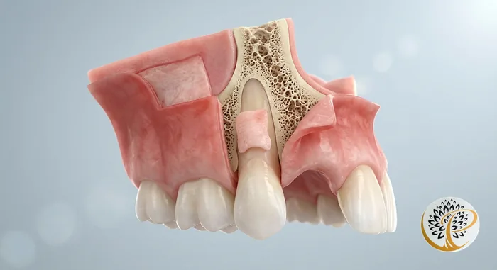
The connective tissue graft consistently produces the best combination of root coverage percentage, color match, and long-term stability across published systematic reviews, which is why it remains the default choice for esthetic recession defects on visible teeth.
When researchers compare graft techniques for treating Miller class one and class two recession defects (the categories most amenable to root coverage), the CTG averages 85 to 95 percent root coverage at one year in well-conducted studies. Free gingival grafts achieve adequate root coverage in some cases but with less reliability and a less natural color blend. Acellular dermal matrix grafts approach CTG outcomes in many studies but with slightly more variability. Pedicle grafts are highly successful when anatomy permits but apply to a narrower set of cases.
The biological reason CTG performs well is that the graft is sandwiched between the patient’s own surface tissue and the underlying bed, which gives it dual blood supply during the critical first week of healing. The covering flap also provides natural color and contour at the gumline, so the cosmetic result blends with surrounding teeth.
The trade-off is the donor site. Harvesting connective tissue from the palate creates a covered donor wound that heals over two to three weeks. Most patients report the palate site as the most noticeable part of recovery, more than the recipient site itself. The discomfort is manageable with over-the-counter pain medication for most patients, and we send everyone home with a custom palatal stent or covering as appropriate.
What Happens During the Procedure Itself?
A typical gum grafting appointment lasts ninety minutes to two hours, is performed under local anesthesia (with sedation available), and the patient leaves with sutures at one or two sites and detailed home care instructions for the first week.
The visit begins with confirmation of the treatment plan, anesthesia, and a final mapping of the recession defects to be treated. The recipient site is prepared first. The root surface is meticulously cleaned and conditioned. A partial-thickness flap is elevated to receive the graft. The donor site is then accessed (palate for autogenous grafts, or no donor site if acellular dermal matrix is being used). The graft is harvested and trimmed to the dimensions of the recipient bed. Sutures secure the graft into position and close the donor area.
Most patients tolerate the procedure well. Patients with significant anxiety can be treated with oral or IV sedation, which is described in our sedation dentistry guide. The actual chair time feels manageable for most patients because the local anesthetic is profound and the work is methodical rather than dramatic.
You leave the office with the surgical sites covered by the sutures themselves rather than by an external dressing in most cases. We provide written instructions, a soft-food guide, and prescriptions or recommendations for any medications appropriate to your case.
What Does the Day-by-Day Recovery Timeline Look Like?
The first three days involve the most swelling and tenderness, days four through seven involve gradual normalization with continued soft-food eating, week two brings substantial healing of the donor site, and the recipient site typically reaches mature healing at twelve to sixteen weeks.
Days one and two are the most cautious window. Some bleeding from the recipient site is normal for the first twenty-four hours and is managed by gentle pressure with damp gauze when needed. Swelling tends to peak around day two or three and is reduced by intermittent cold packs against the cheek (twenty minutes on, twenty minutes off) during the first forty-eight hours. Most patients use over-the-counter ibuprofen or acetaminophen for pain control. Prescription pain medication is rarely necessary but is available if your case warrants it.
Days three through seven feel progressively better. The donor site (if from the palate) is the most prominent source of awareness during this stage because eating creates contact with the healing area. A chlorhexidine rinse is typically prescribed for the first two weeks to control bacterial load without requiring brushing of the surgical sites. Most patients return to office work or school within two to three days; physically demanding work or exercise should wait the full first week.
Week two brings noticeable improvement. The palate begins to feel close to normal and the recipient site looks pink and healthy, with sutures still in place. Sutures are typically removed at the two-week post-operative visit, although many practices now use absorbable sutures that dissolve on their own. The graft itself is firmly attached at this point but the surface tissue is still maturing.
Weeks three through twelve involve cosmetic maturation. The graft tissue, which often looks slightly different in color in the first weeks, blends with surrounding tissue over time. By twelve to sixteen weeks the result is essentially mature, and we evaluate root coverage, tissue thickness, and patient comfort at the final follow-up visit. Some additional cosmetic refinement continues for up to a year as the gumline finds its final architecture.
What Are the Diet and Activity Restrictions?
Soft, cool foods for the first week, no straws, no spitting forcefully, no contact sports for two weeks, and no smoking for as long as the patient is willing because tobacco directly impairs graft healing.
The diet during the first three days centers on cool or room-temperature soft foods. Yogurt, smoothies (eaten with a spoon rather than a straw), pudding, mashed potatoes, scrambled eggs, soft pasta, and similar foods work well. Hot foods and spicy foods irritate the surgical sites and should wait until day three or four. Crunchy foods (chips, nuts, raw vegetables) can disturb the graft and are off-limits for at least one week.
Straws create negative pressure in the mouth that can dislodge clots or strain the graft, so they are avoided for the first week. Forceful spitting after rinsing has the same effect; rinses are gentle, allowing fluid to fall out of the mouth into a sink. Vigorous physical activity raises blood pressure and increases the risk of bleeding from the surgical sites; light activity is fine after a few days but anything strenuous waits a week to ten days.
Smoking deserves a separate mention because it has the largest negative impact on graft success of any modifiable factor. Nicotine constricts the small blood vessels that the graft relies on for survival during the first week, and the published failure rates in active smokers are meaningfully higher than in non-smokers. We have an honest conversation about this with patients who smoke, and we recommend stopping for at least two weeks before and four weeks after the procedure at minimum.
How Do You Care for the Graft Site During the First Month?
Gentle hygiene with no direct brushing of the surgical sites for two weeks, chlorhexidine rinses as prescribed, careful flossing of adjacent untreated teeth, and prompt communication with the office for any signs of infection or graft displacement.
The mouth needs to stay clean during healing, but the graft site itself should not be touched with a toothbrush during the first two weeks. Patients continue to brush all other teeth normally and use a chlorhexidine rinse over the surgical area instead of direct brushing. A soft-bristled toothbrush is reintroduced gently at week two starting with very light pressure on the graft site, and full normal brushing resumes by week three or four as the office advises at follow-up.
Flossing the treated area is suspended until the periodontist clears it, typically at four to six weeks depending on healing. Adjacent teeth that were not treated are flossed normally because biofilm anywhere in the mouth contributes to inflammation that affects healing.
Reasons to call the office include any of the following: increasing rather than decreasing pain after day three, swelling that worsens after day four, discharge with an unpleasant taste, fever, or any sense that the graft has moved or come loose. None of these are common but all are worth a phone call rather than waiting for the scheduled follow-up. Most concerns are easily addressed if caught early.
What Are the Long-Term Success Rates and Outcomes?
Properly performed gum grafts have ten-year success rates above 90 percent in most published studies, and the gain in keratinized tissue and root coverage is generally durable provided the patient maintains good periodontal health and consistent maintenance care.
Success in grafting is measured several ways. The most cited metric is root coverage percentage, meaning how much of the previously exposed root is now covered by gum tissue. CTG procedures average 85 to 95 percent coverage in the literature, with complete coverage achieved in roughly half of well-selected cases. The second metric is the gain in attached, keratinized tissue thickness, which protects the tooth from future recession. The third is patient-reported satisfaction with comfort, sensitivity, and appearance.
Long-term durability depends on what caused the original recession. If the cause was aggressive brushing technique, switching to a soft-bristled brush and a gentler stroke prevents recurrence. If the cause was orthodontic movement of teeth past the boundaries of their bony housing, the graft compensates but the underlying anatomical situation remains, so maintenance matters. If the cause was active periodontitis, the disease must be controlled or the recession will recur regardless of how well the graft healed initially.
Periodontal maintenance every three to four months for patients with a periodontitis history, or every six months for patients without periodontitis, is the framework that preserves grafting outcomes over decades. Patients who follow through with maintenance retain their grafts well. Patients who disappear from regular care are the ones whose results regress.
Ready to Restore Your Gum Health?
If you have been told you need a gum graft and want a clear, in-person evaluation of which technique is appropriate for your situation, the next step is a consultation that includes recession measurement, photography for treatment planning, and a transparent discussion of options, recovery, and cost. Dr. Praveen Parachuru completed his periodontics certificate at the University of Minnesota alongside a PhD in Immunology, which means grafting cases at Prosper Periodontics are evaluated by a clinician trained in both the surgical technique and the tissue biology that drives long-term graft survival. We see patients from Prosper, Frisco, McKinney, Celina, and Aubrey at our office at 2300 E Prosper Trail Suite #20.
To schedule a consultation, call (972) 787-1122 or request an appointment online. Learn more on the gum recession and grafting service page or, for nearby patients, the Frisco gum grafting page covers what to expect for that area specifically. Financing options are available through our Cherry financing partner for patients who want to spread the investment over time.